AAV-GCaMP6s and AAV-DA2h were used to investigate the real-time activity of VTA-DA neurons. AAV-hM3Dq and AAV-hM4Di were used for chemogenetic manipulation. AAV- ChR2 and AAV-NpHR were used for optogenetic manipulation. (All viruses were packaged by
BrainVTA)
The viruses used in this article from BrainVTA are in the table below
|
Calcium sensors |
PT-0091 rAAV2/9-hSyn-DIO-GCaMP6s
PT-0145 rAAV2/9-hSyn-GCaMP6s |
|
Neurotransmitter sensors |
PT1301 rAAV9-hSyn-DA2h |
|
Chemogenetic |
PT-0042 rAAV2/9-EF1a-DIO-hM3Dq-EGFP
PT-0043 rAAV2/9-EF1a-DIO-hM4Di-EGFP |
|
Optogenetic |
PT-0002 rAAV2/9-EF1a-DIO-ChR2-mCherry
PT-0007 rAAV2/9-EF1a-DIO-NpHR-mCherry |
|
CRE Recombinase |
PT-0407 rAAV2/Retro-hSyn-cre-mCherry |
|
Control |
PT-0013 rAAV-Ef1a-DIO-mCherry-WPRE-pA
PT-0012 rAAV-Ef1a-DIO-EGFP-WPRE-pA |
Huan Gui, Chenxi Liu, Haifeng He, Jie Zhang, Hong Chen, Yi Zhang
Pub Date: 2021-04-06,
DOI: 10.3389/fncel.2021.671473,
Email: [email protected]
The role of the dopaminergic pathway in general anesthesia and its potential mechanisms are still unknown. In this study, we used c-Fos staining combined with calcium fiber photometry recording to explore the activity of ventral tegmental area (VTA) dopaminergic neurons (VTA-DA) and nucleus accumbens (NAc) neurons during sevoflurane anesthesia. A genetically encoded dopamine (DA) sensor was used to investigate the function of the NAc in sevoflurane anesthesia. Chemogenetics and optogenetics were used to explore the role of the VTA-DA in sevoflurane anesthesia. Electroencephalogram (EEG) spectra, time of loss of righting reflex (LORR) and recovery of righting reflex (RORR) were recorded as assessment indicators. We found that VTA-DA and NAc neurons were inhibited during the induction period and were activated during the recovery period of sevoflurane anesthesia. The fluorescence signals of dopamine decreased in the induction of and increased in the emergence from sevoflurane anesthesia. Activation of VTA-DA and the VTA
DA-NAc pathway delayed the induction and facilitated the emergence, accompanying with the reduction of delta band and the augment of gamma band. These data demonstrate that VTA-DA neurons play a critical role in modulating sevoflurane anesthesia via the VTA
DA-NAc pathway.
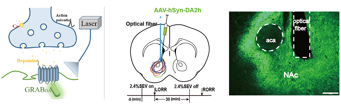 Figure 1. Dynamics of extracellular dopamine in the nucleus accumbens (NAc) in response to sevoflurane anesthesia.
The role of the dopaminergic pathway in general anesthesia and its potential mechanisms are still unknown. Using c-Fos staining combined with calcium fiber photometry recording, the authors explored the activity of ventral tegmental area (VTA) dopaminergic neurons (VTA-DA) and nucleus accumbens (NAc) neurons during sevoflurane anesthesia. The results demonstrated that VTA-DA neurons play a critical role in modulating sevoflurane anesthesia via the VTADA-NAc pathway.
BrainVTA offers viral vector construction & virus packaging services for AAV, LV, RABV, PRV, HSV and VSV that help researchers explore questions about genes, neurons, circuitry structure, function of brain network, mechanism and treatment of diseases.
If you have any needs, just email us at
[email protected].


