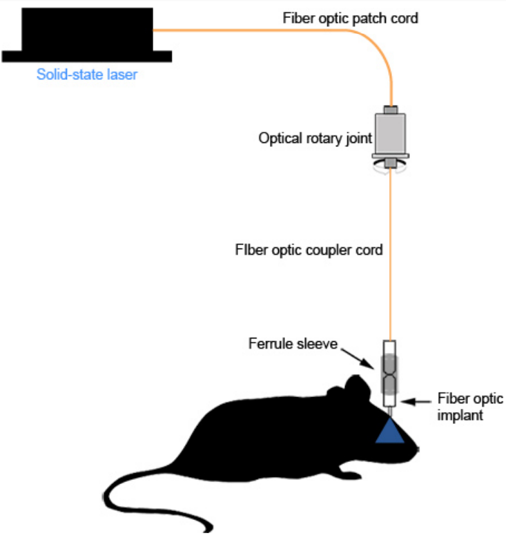
Fiber photometry is widely used to monitor neuronal activity in freely-behaving animals in modern experimental neuroscience research. Fiber photometry techniques for neuroscience employ an optical fiber implanted into the brain to enable study of the function of specific populations of neurons. Take the injection of ChR2 virus into VLO as the example, the detailed procedures of optical fiber implantation are as follows.
 kevin et al., jove, 2012.
STEP 1:
Vglut2-IRES-cre mice injected with pAAV-Efla-DIO-hChR2(H134R)-mCherry virus into bilateral VLO and Vgat-IRES- cre mice injected with pAOV-EF1a-DIO-MAC-mCherry virus into bilateral VLO (and corresponding control mice) were returned to the cage for one week, then slowly implant optical fiber to the brain region of interest.
STEP 2:
First anesthetize the animal, expose the skull and brain tissue as described in the process of virus injection.
STEP 3:
Insert a customized LED (Φ200 μm, numerical aperture NA 0.73, long 2.4 mm , diameter 2.0 mm) with wireless bipolar fiber into the target position (AP = +2.5 mm, ML = ±1.0 mm, DV =-1.5 mm), the depth is 0.3 mm higher than the virus injection site to ensure that the light covers a large enough area. In addition, choose two asymmetric positions on the brain plane to fix two stainless steel screws and fill the gaps around the optical fiber and the screws with tissue adhesive, and finally fix the LED with dental cement.
If you have any questions,please email us at
[email protected].


