AAV-hM3D was used for chemogenetics manipulation. (From
BrainVTA)
The viruses used in this article from BrainVTA are in the table below
|
Chemogenetics |
AAV-EF1α-DIO-hM3D-mCherry |
Lin Chen, Jun Hu, Jiankun Mu, Chao Li, Guang-Yan Wu, Chao He, Youhong Xie, Jian-Ning Ye
Pub Date: 2021-08-03,
DOI: 10.1016/j.bbr.2021.113511,
Email: [email protected]
Prefrontal ischemia can cause impairments in learning and memory, executive functions and cognitive flexibility. However, the related cellular mechanisms at the early stage are still elusive. The present study used ischemic stroke in medial prefrontal cortex and systemically investigated the electrophysiological changes of the parvalbumin (PV+) interneurons 12 h post ischemia. We found that Ih and the related voltage sags in PV+ interneurons are downregulated post ischemia, which correlates with hyperpolarization of the membrane potentials and increased input resistance in these interneurons. Consistent with the suppression of Ih, postischemic PV+ interneurons exhibited a reduction in excitability and exerted a less inhibitory control over the neighboring pyramidal excitatory neurons. Moreover, we found that specifically chemogenetic activation of PV+ neurons at early stage ameliorated prefrontal ischemia-induced spatial working memory dysfunction in T-maze without effects on the locomotor coordination and balance. In contrast, suppression of PV+ neurons by blockade of Ih leaded to further aggravate the damage of spatial memory. These findings indicate that dysfunctional Ih in the PV+ neuron postischemia induces the imbalance of excitation and inhibition, which might represent a novel mechanism underlying the prefrontal ischemia-induced cognitive impairment.
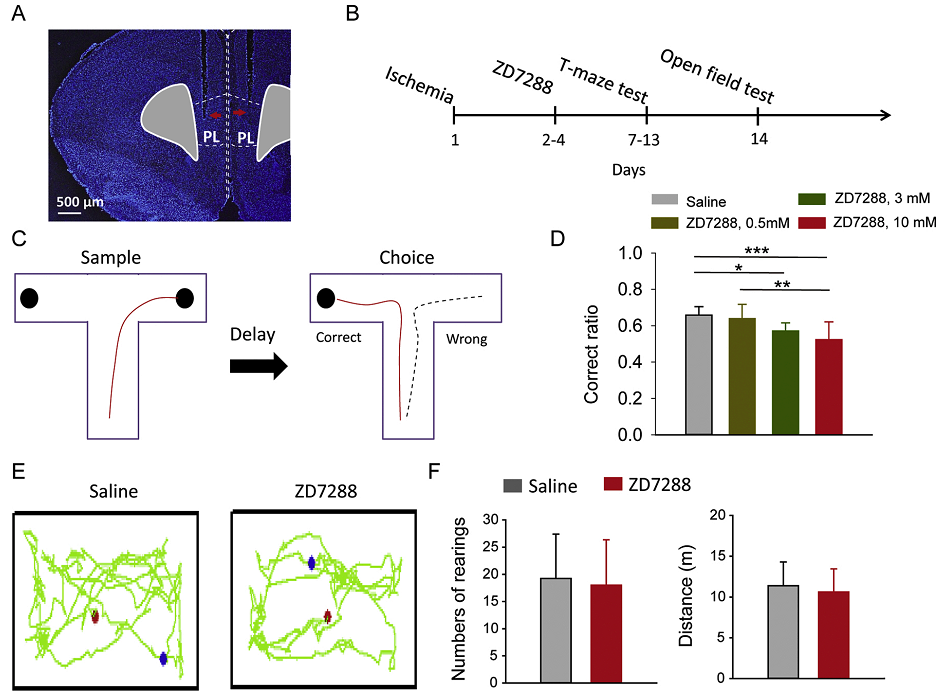 Figure 1. Suppression of PV+ neurons by blockade of Ih aggravated the damage of spatial memory.
In this study, the authors used animal model of mPFC ischemic stroke and investigated how the electrophysiological properties of interneurons are altered following 12 h post stroke, during which the stroke-induced cellular dysfunction is prominent. The findings indicate that dysfunctional Ih in the PV+ neuron postischemia induces the imbalance of excitation and inhibition, which might represent a novel mechanism underlying the prefrontal ischemia-induced cognitive impairment.
BrainVTA offers viral vector construction & virus packaging services for AAV, LV, RABV, PRV, HSV and VSV that help researchers explore questions about genes, neurons, circuitry structure, function of brain network, mechanism and treatment of diseases.
If you have any needs, just email us at
[email protected].


