AAV-GCaMP6m was used for fiber photometry experiment. (From
BrainVTA)
The viruses used in this article from BrainVTA are in the table below
|
Calcium sensors |
AAV-Ef1α-DIO-GCaMP6m-eYFP |
|
Control |
PT-0012 AAV-Ef1α-DIO-eYFP |
Zhijun Diao, Li Yao, Qiangqiang Cheng, Meilin Wu, Yuanyuan Di, Zhaoqiang Qian, Chunling Wei, Yingxun Liu, Yingfang Tian, Wei Ren
Pub Date: 2022-08-13,
DOI: 10.1007/s12035-021-02515-6,
Email: [email protected]
The activity of the midbrain dopamine system reflects the valence of environmental events and modulates various brain structures to modify an organism’s behavior. A series of recent studies reported that the direct and indirect pathways in the striatum are critical for instrumental learning, but the dynamic changes in dopamine neuron activity that occur during negative reinforcement learning are still largely unclear. In the present study, by using a negative reinforcement learning paradigm employing foot shocks as aversive stimuli, bidirectional changes in substantia nigra pars compacta (SNc) dopamine neuron activity in the learning and habituation phases were observed. The results showed that in the learning phase, before mice had mastered the skill of escaping foot shocks, the presence of foot shocks induced a transient reduction in the activity of SNc dopamine neurons; however, in the habituation phase, in which the learned skill was automated, it induced a transient increase. Microinjection of a dopamine D1 receptor (D1R) or D2 receptor (D2R) antagonist into the dorsomedial striatum (DMS) significantly impaired learning behavior, suggesting that the modulatory effects of dopamine on both the direct and indirect pathways are required. Moreover, during the learning phase, excitatory synaptic transmission to DMS D2R-expressing medium spiny neurons (D2-MSNs) was potentiated. However, upon completion of the learning and habituation phases, the synapses onto D1R-expressing medium spiny neurons (D1-MSNs) were potentiated, and those onto D2-MSNs were restored to normal levels. The bidirectional changes in both SNc dopamine neuron activity and DMS synaptic plasticity might be the critical neural correlates for negative reinforcement learning.
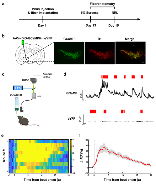 Figure 1. Calcium signals recording during licking sucrose in mice expressing GCaMP before negative reinforcement learning.
The present study investigated the dynamic changes in SNc dopamine neuron activity during the negative reinforcement learning process. The resulting neural plastic changes in DMS direct and indirect pathway neurons were also observed in the learning and habituation phases of the negative reinforcement learning process.
BrainVTA offers viral vector construction & virus packaging services for AAV, LV, RABV, PRV, HSV and VSV that help researchers explore questions about genes, neurons, circuitry structure, function of brain network, mechanism and treatment of diseases.
If you have any needs, just email us at
[email protected].


