Rabies-virus system was used for tracing of afferent inputs onto SON
AVP and PVN
AVP neurons. (From
BrainVTA)
The viruses used in this article from BrainVTA are in the table below
|
Tracing Helper |
PT-0062 AAV-DIO-EGFP-2A-TVA
PT-0023 AAV-DIO-RG, |
|
RV |
R01002 RV-SAD19-DG-dsRed (EnvA) |
Hao-Hua Wei, Xiang-Shan Yuan, Ze-Ka Chen, Pei-Pei Chen, Zhe Xiang, Wei-Min Qu, Rui-Xi Li, Guo-Min Zhou, Zhi-Li Huang
Pub Date: 2021-06-15,
DOI: 10.1016/j.expneurol.2021.113784,
Email: [email protected]
Arginine vasopressin (AVP) neurons in the hypothalamic supraoptic nucleus (SON) and paraventricular nucleus (PVN) are involved in important physiological behaviors, such as controling osmotic stability and thermoregulation. However, the presynaptic input patterns governing AVP neurons have remained poorly understood due to their heterogeneity, as well as intermingling of AVP neurons with other neurons both in the SON and PVN. In the present study, we employed a retrograde modified rabies-virus system to reveal the brain areas that provide specific inputs to AVP neurons in the SON and PVN. We found that AVP neurons of the SON and PVN received similar input patterns from multiple areas of the brain, particularly massive afferent inputs from the diencephalon and other brain regions of the limbic system; however, PVNAVP neurons received relatively broader and denser inputs compared to SONAVP neurons. Additionally, SONAVP neurons received more projections from the median preoptic nucleus and organum vasculosum of the lamina terminalis (a circumventricular organ), compared to PVNAVP neurons, while PVNAVP neurons received more afferent inputs from the bed nucleus of stria terminalis and dorsomedial nucleus of the hypothalamus, both of which are thermoregulatory nuclei, compared to those of SONAVP neurons. In addition, both SONAVP and PVNAVP neurons received direct afferent projections from the bilateral suprachiasmatic nucleus, which is the master regulator of circadian rhythms and is concomitantly responsible for fluctuations in AVP levels. Taken together, our present results provide a comprehensive understanding of the specific afferent framework of AVP neurons both in the SON and PVN, and lay the foundation for further dissecting the diverse roles of SONAVP and PVNAVP neurons.
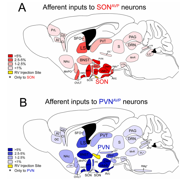 Figure 1. Summary of the major afferent inputs onto SON and PVNAVP neurons. Brain regions that afferently project onto AVP neurons in the SON.
In this study, the authors employed a modified rabies virus and Cre-dependent adeno-associated viruses (AAVs) in AVP-Cre mice to map presynaptic afferent inputs onto SONAVP and PVNAVP neurons. Collectively, these findings reveal differential presynaptic input patterns onto SONAVP and PVNAVP neurons, and provide a neuroanatomic foundation for further studies interrogating the neural circuits that underlie diverse physiological functions of SONAVP and PVNAVP neurons.
BrainVTA offers viral vector construction & virus packaging services for AAV, LV, RABV, PRV, HSV and VSV that help researchers explore questions about genes, neurons, circuitry structure, function of brain network, mechanism and treatment of diseases.
If you have any needs, just email us at
[email protected].


