AAV-Ferritin was used for gene over-expression. (From
BrainVTA)
The viruses used in this article from BrainVTA are in the table below
|
Custom-Made AAV |
rAAV2-retro-CAG-Ferritin |
|
Control |
rAAV2-retro-CAG-EGFP |
Aoling Cai, Ning Zheng, Garth J Thompson, Yang Wu, Binbin Nie, Kunzhang Lin, Peng Su, Jinfeng Wu, Anne Manyande, LingQiang Zhu, Jie Wang, Fuqiang Xu
Pub Date: 2021-07-20,
DOI: 10.1002/hbm.25596,
Email: [email protected]
The investigation of neural circuits is important for interpreting both healthy brain function and psychiatric disorders. Currently, the architecture of neural circuits is always investigated with fluorescent protein encoding neurotropic virus and ex vivo fluorescent imaging technology. However, it is difficult to obtain a whole-brain neural circuit connection in living animals, due to the limited fluorescent imaging depth. Herein, the noninvasive, whole-brain imaging technique of MRI and the hypotoxicity virus vector AAV (adeno-associated virus) were combined to investigate the whole-brain neural circuits in vivo. AAV2-retro are an artificially-evolved virus vector that permits access to the terminal of neurons and retrograde transport to their cell bodies. By expressing the ferritin protein which could accumulate iron ions and influence the MRI contrast, the neurotropic virus can cause MRI signal changes in the infected regions. For mice injected with the ferritin-encoding virus vector (rAAV2-retro-CAG-Ferritin) in the caudate putamen (CPu), several regions showed significant changes in MRI contrasts, such as PFC (prefrontal cortex), HIP (hippocampus), Ins (insular cortex) and BLA (basolateral amygdala). The expression of ferritin in those regions was also verified with ex vivo fluorescence imaging. In addition, we demonstrated that changes in T2 relaxation time could be used to identify the spread area of the virus in the brain over time. Thus, the neural connections could be longitudinally detected with the in vivo MRI method. This novel technique could be utilized to observe the viral infection process and detect the neural circuits in a living animal.
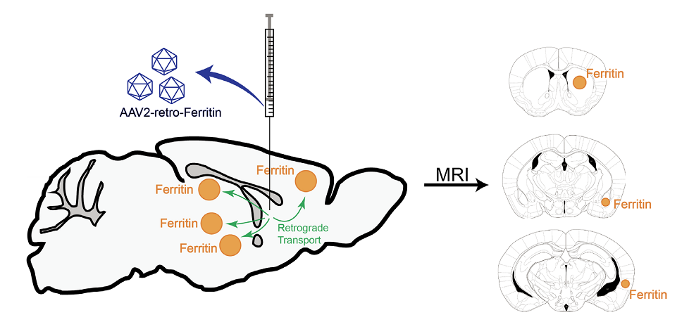 Figure 1. Illustration of the schedule of the experiment.
In this study, the authors developed a novel tool for in vivo whole-brain neural network imaging. They loaded the ferritin gene onto a retrograde transporting AAV vector, delivered it to the caudate-putamen (CPu) of mice and imaged these mice with in vivo MRI. In doing so, we were able to visualize a CPu-connected network that includes the upstream brain regions sending projection to the CPu. The ferritin-encoding retrograde transporting AAV vector enabled the investigation of neural network in living animals and long-term observation of the virus infection.
BrainVTA offers viral vector construction & virus packaging services for AAV, LV, RABV, PRV, HSV and VSV that help researchers explore questions about genes, neurons, circuitry structure, function of brain network, mechanism and treatment of diseases.
If you have any needs, just email us at
[email protected].


