(RV)-ΔG-GFP was used to inject into the NDB region for cell counting. (From
BrainVTA)
The viruses used in this article are in the table below
Qiuyuan Zhong, Anan Li, Rui Jin, Dejie Zhang, Xiangning Li, Xueyan Jia, Zhangheng Ding, Pan Luo, Can Zhou, Chenyu Jiang, Zhao Feng, Zhihong Zhang, Hui Gong, Jing Yuan, Qingming Luo
Pub Date: 2021-03-01,
DOI: 10.1038/s41592-021-01074-x,
Email: [email protected]
The microscopic visualization of large-scale three-dimensional (3D) samples by optical microscopy requires overcoming challenges in imaging quality and speed and in big data acquisition and management. We report a line-illumination modulation (LiMo) technique for imaging thick tissues with high throughput and low background. Combining LiMo with thin tissue sectioning, we further develop a high-definition fluorescent micro-optical sectioning tomography (HD-fMOST) method that features an average signal-to-noise ratio of 110, leading to substantial improvement in neuronal morphology reconstruction. We achieve a >30-fold lossless data compression at a voxel resolution of 0.32 × 0.32 × 1.00 μm3, enabling online data storage to a USB drive or in the cloud, and high-precision (95% accuracy) brain-wide 3D cell counting in real time. These results highlight the potential of HD-fMOST to facilitate large-scale acquisition and analysis of whole-brain high-resolution datasets.
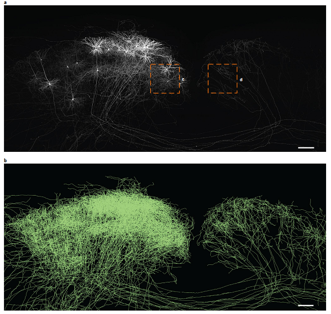 Figure 1. Automatic signal segmentation on a raw data block with 11,200 × 5,200 × 3,200 voxels.
In this study, the researchers report a line-illumination modulation (LiMo) technique for imaging thick tissues with high throughput and low background, combining LiMo with thin tissue section-ing, they further develop a high-definition fluorescent micro-optical sectioning tomography (HD-fMOST) method. This allows for acquisition of more information about neuronal morphology than previously possible.
BrainVTA offers viral vector construction & virus packaging services for AAV, LV, RABV, PRV, HSV and VSV that help researchers explore questions about genes, neurons, circuitry structure, function of brain network, mechanism and treatment of diseases.
If you have any needs, just email us at
[email protected].


