AAVs of tracing helper and RV were used for retrograde monosynaptic tracing.
The viruses used in this article are in the table below
|
Tracing Helper |
AAV-DIO-EGFP–TVA
AAV-DIO-RG |
|
Control |
AAV-DIO-EmGFP |
|
RV |
SADΔG-mCherry |
Qingchun Guo, DaqingWang, Xiaobin He, Qiru Feng, Rui Lin, Fuqiang Xu, Ling Fu, Minmin Luo
Pub Date: 2015-04-01,
DOI: 10.1371/journal.pone.0123381,
Email: [email protected]
The dorsal striatum integrates inputs from multiple brain areas to coordinate voluntary movements, associative plasticity, and reinforcement learning. Its projection neurons consist of the GABAergic medium spiny neurons (MSNs) that express dopamine receptor type 1 (D1) or dopamine receptor type 2 (D2). Cholinergic interneurons account for a small portion of striatal neuron populations, but they play important roles in striatal functions by synapsing onto the MSNs and other local interneurons. By combining the modified rabies virus with specific Cre- mouse lines, a recent study mapped the monosynaptic input patterns to MSNs. Because only a small number of extrastriatal neurons were labeled in the prior study, it is important to reexamine the input patterns of MSNs with higher labeling efficiency. Additionally, the whole-brain innervation pattern of cholinergic interneurons remains unknown. Using the rabies virus-based transsynaptic tracing method in this study, we comprehensively charted the brain areas that provide direct inputs to D1-MSNs, D2-MSNs, and cholinergic interneurons in the dorsal striatum.We found that both types of projection neurons and the cholinergic interneurons receive extensive inputs from discrete brain areas in the cortex, thalamus, amygdala, and other subcortical areas, several of which were not reported in the previous study. The MSNs and cholinergic interneurons share largely common inputs from areas outside the striatum. However, innervations within the dorsal striatum represent a significantly larger proportion of total inputs for cholinergic interneurons than for the MSNs. The comprehensive maps of direct inputs to striatal MSNs and cholinergic interneurons shall assist future functional dissection of the striatal circuits.
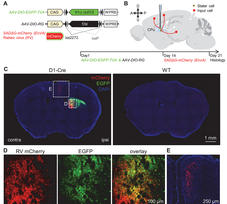 Figure 1. The method of rabies-based retrograde tracing.
In this study, the authors studied the whole-brain input patterns of D1-and D2-MSNs and cholinergic interneurons by selectively infecting these neurons with the modified rabies virus. The results reveal that the two types of MSNs and the cholinergic interneurons receive extensive inputs from discrete areas in the cortex, thalamus, amygdala, globus pallidus, and SNc. Moreover, cholinergic interneurons receive particularly dense local innervations from non-cholinergic neurons within the dorsal striatum.
BrainVTA offers viral vector construction & virus packaging services for AAV, LV, RABV, PRV, HSV and VSV that help researchers explore questions about genes, neurons, circuitry structure, function of brain network, mechanism and treatment of diseases.
If you have any needs, just email us at
[email protected].


