AAVs of tracing helper and RV were used for retrograde monosynaptic tracing. PRV was used to map the multi-synaptic input connections. RV was used to study the whole-brain organizations of mono-synaptic inputs to the D1R- or D2R-MSNs in the medial part of the OT. (All the viruses were from
BrainVTA)
The viruses used from BrainVTA in this article are in the table below
|
Tracing Helper |
AAV9-EF1a-Dio-GFP-TVA
AAV9-EF1a-DiO-RV-G |
|
Control |
AAV9-EF1a-floxed-EGFP |
|
PRV |
PRV-152 |
|
RV |
RV-EnvA-1G-dsRed |
Zhijian Zhang, Hongruo Zhang, Pengjie Wen, Xutao Zhu, Li Wang, Qing Liu, Jie Wang, Xiaobin He, Huadong Wang, Fuqiang Xu
Pub Date: 2017-08-15,
DOI: 10.3389/fncir.2017.00052,
Email: [email protected]
The medial part of the olfactory tubercle (OT) is a brain structure located at the interface of the reward and olfactory system. It is closely related to pheromone-rewards, natural reinforcement, addiction and many other behaviors. However, the structure of the anatomic circuitry of the medial part of the OT is still unclear. In the present study, the medial part of the OT was found to be highly connected with a wide range of brain areas with the help of the pseudorabies virus tracing tool. In order to further investigate the detailed connections for specific neurons, another tracing tool-rabies virus was utilized for D1R-cre and D2R-cre mice. The D1R and D2R neurons in the medial part of the OT were both preferentially innervated by the olfactory areas, especially the piriform cortex, and both had similar direct input patterns. With the help of the adeno-associated virus labeling, it was found that the two subpopulations of neurons primarily innervate with the reward related brain regions, with slightly less axons projecting to the olfactory areas. Thus, the whole-brain input and output circuitry structures for specific types of neurons in the medial part of the OT were systematically investigated, and the results revealed many unique connecting features. This work could provide new insights for further study into the physiological functions of the medial part of the OT.
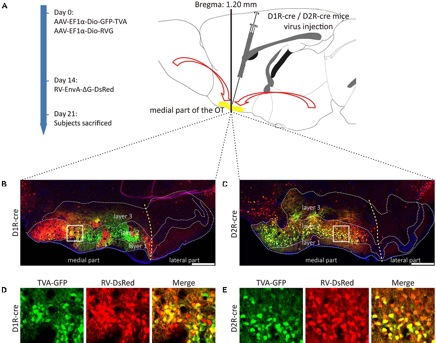 Figure 1. Rabies virus (RV) trans-monosynaptic tracing from the D1R- and D2R-MSNs of the medial part of the OT.
Here, the aim was to use a combination of viral toolboxes to reveal the circuitry structure of the medial part of the OT. In this study, the multi-synaptic input connections were mapped with the pseudorabies virus (PRV). Furthermore, the whole-brain organizations of mono-synaptic inputs to the D1R- or D2R-MSNs in the medial part of the OT were studied, utilizing the modified rabies virus (RV). At last, the global axonal efferent patterns of these two types of neurons were examined with adeno-associated virus (AAV).
BrainVTA offers viral vector construction & virus packaging services for AAV, LV, RABV, PRV, HSV and VSV that help researchers explore questions about genes, neurons, circuitry structure, function of brain network, mechanism and treatment of diseases.
If you have any needs, just email us at
[email protected].


