AAV-s100a4 was used to overexpress mice S100A4. (From
BrainVTA)
The viruses used in this article from BrainVTA are in the table below
|
Custom-Made AAVs |
rAAV-Eflα-s100a4-EGFP-WPRE |
|
Control |
rAAV-Eflα-EGFP-WPRE-Pa |
Jiayi Yang, Ning Yang, Jinyuan Luo, Gumeng Cheng, Xiao Zhang, Tao He, Yiqiao Xing
Pub Date: 2020-10-06,
DOI: 10.1016/j.exer.2020.108281,
Email: [email protected]
Background:Glaucoma is characterized by the neurodegeneration of retinal ganglion cells (RGCs) and the optic nerve. Numerous studies have reported that S100A4 participates in the metastasis of tumor cells and nerve protection. This study was intended to explore the role of S100A4 on RGCs under retinal ischemia-reperfusion (I/R) injury in mice.
Methods:C57BL/6J mice were used to induce retinal I/R injury. The intravitreal administration of rAAV-EF1α-s100a4-EGFP-WPRE (rAAV-S100A4) or rAAV-EF1α-EGFP-WPRE-Pa was performed 4 weeks before I/R injury. Expression of S100A4 was detected by quantitative real-time PCR, immunofluorescence staining of retinal sections and western blot. Surviving RGCs were quantified using immunofluorescence staining. Staining of TUNEL was utilized to evaluate the apoptosis of retinal cells. Electroretinogram (ERG) was used to analyze retinal function. Expression of Akt, phospho-Akt, Bcl-2, and Bax were determined using western blotting to investigate the potential mechanisms of S100A4.
Results:Retinal S100A4 level had no statistical difference 7 days after I/R injury. The rAAV-S100A4 was clearly demonstrated by the green fluorescence protein in many layers of the retina after intravitreal injection and up-regulated the expression of S100A4. I/R injury resulted in an increase of the apoptosis of retinal cells and the reduction of surviving RGCs, however, overexpressed S100A4 inhibited the apoptosis of cells and a decrease of RGCs. ERG analysis showed a drop on amplitude of a-wave and b-wave was impeded to some extent by overexpressing of S100A4. Up-regulation of S100A4 raised the expression of phospho-Akt and reduced Bax expression. Nevertheless, there were no significant changes in the levels of Bcl-2 and total Akt.
Conclusion:Our results indicate the neuroprotective effects of overexpressed S100A4 on RGCs by activating the Akt pathway and then inhibiting the apoptosis of cells after I/R injury. The use of S100A4 protein may be a novel therapeutic strategy for glaucoma.
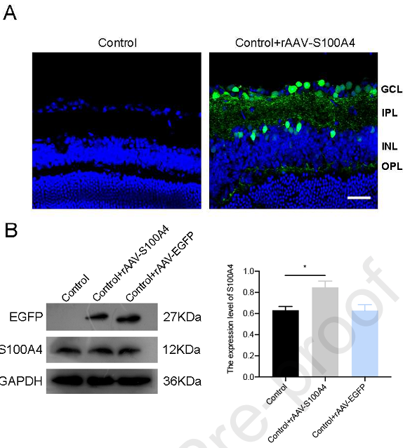 Figure 1. The rAAV-Eflα-s100a4-EGFP-WPRE transferred into mice retinas successfully and up-regulated the expression level of S100A4
This study was aimed to explore the role of S100A4 on RGCs under retinal ischemia-reperfusion (I/R) injury in mice. In this study, the authors used a mouse model of retinal ischemia-reperfusion (I/R) to simulate the death of RGCs in glaucoma and to study the effect of S100A4 on RGCs. Our results indicated that the neuroprotective effects of overexpressed S100A4 on RGCs by activating Akt pathway and then inhibiting the apoptosis of cells after I/R injury. S100A4 protein may be a novel therapeutic target for glaucoma
BrainVTA offers viral vector construction & virus packaging services for AAV, LV, RABV, PRV, HSV and VSV that help researchers explore questions about genes, neurons, circuitry structure, function of brain network, mechanism and treatment of diseases.
If you have any needs, just email us at
[email protected].


